Sunday, April 28, 2019

PKD Modeling with Kidney Organiods, Kidney Pain Signs, Medicare Rules Modification Could Lead to Dialysis Care Innovation, Kidney Delivery by Drone

Living with PKD

From Medical Daily, By Leian Naduma

Kidney Pain Signs, Symptoms And Causes

Renal or kidney pain is associated with several malfunctions of the organs or recurring infection. Sometimes, it is a mere urinary tract infection that can be remedied by lifestyle changes or prescription medication. Other times, it is a symptom of a serious mental condition. Here are the most common signs, symptoms and causes to help you identify if what you are going through is a mild or serious case of kidney pain.

Your kidneys are bean-shaped organs located on either side of your spine. They filter the blood and balance the number of fluids and electrolytes in your body. The level of pain you experience also depends on whether the cause is prerenal or related to another organ located near it, intrinsic or caused by the kidneys themselves or postrenal, which is typically due to an obstruction below the organs. Thus, the common triggers of kidney pain are an infection, obstruction, growth or trauma, as per VeryWell Health.

Trauma

Kidneys are placed at a vulnerable position in the abdomen which may easily be affected by a blunt force impact or a penetrating wound. If you have abdominal injuries, there is a 10 percent chance that you will also sustain damage to your kidneys. Events such as physical assaults and vehicular accidents result in renal trauma. You can distinguish this cause from others by pressing the kidney area and note a painful sensation. It may also be associated with fever, hematuria, urinary retention, rapid heart rate, decreased alertness and abdominal swelling. These symptoms require emergency treatment.

Renal Obstruction

This is a result of urinary blockage and it is an intrinsic type of kidney pain. The renal obstruction causes unilateral or bilateral pain due to affected ureters. This type of pain is also called obstructive uropathy which is caused by kidney stones, bladder stones, urinary tract infection, an enlarged prostate, pregnancy, long-term catheterization, a blood clot in the kidney, nerve-related bladder weakness, cancer, or vesicoureteral reflux. These cause your kidneys to swell and is referred to as hydronephrosis. You are most likely to experience pain in the groin, flak or abdominal area and may be associated with urinary urgency, fever, nausea and dysuria. Kidney stones cause higher pain levels while the others are gradual when left untreated.

Growth or Cysts

Renal tumors also cause pain and its intensity depends on the growth. The three most common growth abnormalities are renal adenoma, renal cell carcinoma and polycystic kidney disease (PKD). Larger ones do not cause pain until they disturb the architecture of the kidney. When this happens, the pain is persistent and worsens over time. If the tumor is cancerous, you may also experience unexplained weight loss, which suggests an advanced malignancy. PKD is also associated with headaches, high blood pressure, abdominal pain and swelling, hematuria and renal failure.

Infection

Kidney infection is treated with prescription antibiotics. This is caused by viral or fungal exposure that adversely affects the body’s immune system. Patients who have advanced HIV or underwent organ transplants are more vulnerable to this condition.

Prevention And Cure

As per Medicine.Net, most kidney pains can be relieved by drugs like ibuprofen, ketorolac and acetaminophen, which are common medications for pain. Antibiotics are recommended by doctors in infections while those who have kidney stones may need surgery.

Kidney pains caused by infections can be prevented by drinking sufficient amounts of water per day, wearing loose pants, following proper hygiene and refraining from holding your urine for long periods. Kidney stones and abdominal cancers, on the other hand, may be prevented by eating a healthy diet and avoiding salty and fatty foods.


From Health Care Finance, by Jeff Lagasse, Associate Editor

Modifications to Medicare rules could support care innovation for dialysis

Medicare spends approximately $35 billion annually on care for beneficiaries with end-stage renal disease.

In a commentary published in the American Journal of Kidney Diseases, public health researchers suggest adjustments to recently proposed rule changes on how Medicare pays for dialysis services.

Medicare spends approximately $35 billion annually on care for beneficiaries with end-stage renal disease, or kidney failure. That's more than 7 percent of Medicare's total paid claims. Over half a million people receive regular dialysis treatments to manage this condition, with treatment costs averaging about $85,000 a year, according to the study.

IMPACT

Rule changes were proposed about a year ago that would limit the number of dialysis treatments per week that would be paid for by Medicare. Interest groups including nephrologists and patients themselves were concerned that this would limit patient access to innovative treatment options, such as frequent hemodialysis.

Under the current system, Medicare covers three hemodialysis treatments weekly per patient, but it will often pay for additional treatments when the treating nephrologist provides sufficient medical justification.

The recently proposed rule changes would limit such additional payments to exceptional circumstances -- for example, patients with temporary, acute kidney treatment needs. Although nephrologists would not be prevented from providing any "extra" treatments they believe are needed, they would typically bear the costs of doing so.

The researchers discussed the limitations of the current evidence on frequent dialysis treatment, which to date has yielded mixed conclusions. Their suggested changes to Medicare's dialysis payment system were designed to account for these limitations and give Medicare the flexibility to further modify the system in the future as new evidence comes to light.

Under their main proposal, Medicare would establish a new, separate prospective payment system for frequent hemodialysis treatment.

In that way, they said, nephrologists would have greater clarity about how their dialysis care would be paid for, and free them up to pinpoint better ways to treat their patients.

THE TREND

In the first three years of Medicaid expansion due to the Affordable Care Act, the number of patients with end-stage kidney disease who died within a year of starting dialysis decreased in states that expanded Medicaid, compared to non-expansion states, found research published in October.

The adjusted absolute reduction in mortality in expansion states versus non-expansion states was 0.6 percentage points. Since end-stage renal disease affects more than 100,000 Americans each year, 0.6 percentage points equals hundreds of deaths annually.


PKD Research

Select Science

Modeling Kidney Disease with Bioengineered Kidney Organoids

 Dr. Benjamin Freedman, Assistant Professor at the University of Washington

Kidney organoid pioneer, Dr. Benjamin Freedman, explains the bioengineering, generation and application of 3D cell culture in studying kidney disease.

Kidney disease, prevalent in about 14% of the American population, lacks much-needed early interventions to prevent disease progression. Current methods that address kidney disease in a more chronic stage endure additional complexities: for example, artificial kidney devices don’t function as well as a human kidney, and renal transplant procedures need the support of anti-rejection medication.

In this article, we interview Dr. Benjamin Freedman, Assistant Professor at the University of Washington, whose goal is to understand kidney disease in its early stages. “In the medical practice, there's a lot of focus on the end stages of kidney disease and on managing that chronic disease. We are more interested in the early stages and all the different things that can go wrong early on,” says Freedman.

A pioneer in the field of kidney organoids, Freedman chose the 3D cell culture system to study kidney disease. “Organoids are great for this because they can show signs of disease,” Freedman explains. “We can then intervene by treating them with any compound that we're interested in.” A robust model for mimicking disease in vitro, kidney organoids didn’t even exist until a few years ago.

Freedman was the first scientist in the western hemisphere to generate a kidney organoid from pluripotent stem cells. “That was a very exciting moment,” Freedman recalls. “As soon as I saw these structures, I knew that there was something interesting about them. I had all the tools at my disposal to test whether they were indeed kidney.”

And they were.

Bioengineering kidney organoids

Developing and maintaining kidney organoids requires bioengineering capabilities. While the organoids grow and self-assemble from stem cells, their in vitro development deprives them of the vascular perfusion otherwise available in vivo. Tiny microfluidic tubules pass through the kidneys in our bodies, enabling the inlet and outlet of fluid, a resource that kidney organoids grown on a dish don’t receive.

“The kidneys in the body get about 20–25% of the cardiac output at any time point,” explains Freedman. “One thing we're missing in an organoid structure is the ability to perfuse the organoids and the tiny tubules.”

Freedman’s lab has a solution to this problem. “We're trying to make the kidney organoid 2.0, which will incorporate not just the stem cells and their natural ability to form the structures but will also impose a bioengineering design on top of those structures to enable them to really form the very complex types of functional tubules that are found in the body.”

Kidney Organoids in the Freedman lab
Kidney organoids cultured in the Freedman lab. Images from left to right; blood vessels joining kidney organoids; grown from reprogrammed skin cells; showing cyst growth from tubules; kidney podocytes. Image courtesy of the Freedman lab.

Mimicking in vivo environments

The kidney organoid 2.0 grows on a kidney-on-a-chip device incorporated with extracellular matrix in scaffolded arrangements. By seeding the stem cell-derived kidney cells on this matrix-coated chip, the cells can be grown into controlled shapes and arrangements. “It's a fusion between stem cell and bioengineering fields,” explains Freedman. “The chip, about the size of a credit card, has a tiny tubule structure through which we can perfuse liquid. We can then observe the absorption of solutes such as glucose and ions and see how they're transported from one side of the tubule to the other side.”

The procedure to generate organoids uses a thin layer of Corning® Matrigel® matrix at the bottom of the tissue culture dish. The cells are plated on top of this layer, followed by a second thin layer of Matrigel. “This enables the cells to fold up and create more three-dimensional types of structures which are about 200 microns in diameter,” says Freedman. “These contain all the key lineages of the kidney tissue that we're interested in.”

The Matrigel matrix forms a crucial component of the kidney organoid generation process from pluripotent stem cells. “Matrigel matrix helps the cells to survive. They need a malleable support that they can grow into and form more three-dimensional structures in; something that they can actually change and remodel as they're growing,” Freedman explains.

For the kidney-on-a-chip protocol, the Freedman lab uses a relatively strong and stiff extracellular matrix. “We use Corning Collagen I, a highly concentrated form of collagen because these gels need to be stiff for the cells to take on the right shape that we’re interested in,” says Freedman. “If it’s too soft, the whole thing just falls apart, especially because we have fluid running through it.”

Alternatively, after the organoids have formed, they are placed inside larger Corning Collagen I droplets to stimulate the cells to grow out and populate. “This way, we study the ability of these cells to migrate out of the organoid, which has ramifications for certain disease processes,” says Freedman. “The tubular cells, which are normally sedentary, can actually migrate out when you give them an environment like that.”

Better detached than attached

One of the projects in the Freedman lab involves studying polycystic kidney disease using the kidney organoid model. “In polycystic kidney disease, the tubules, which are normally very narrow, swell up to form large balloon-life structures. They eventually crowd out and destroy all the healthy kidney tissue,” explains Freedman. “We’ve been able to get this to actually happen in our kidney organoids by mutating a gene in the stem cells and derive organoids that are mutants. The organoids swell just like the kidney tubules in the disease.”

This swelling process, however, depends on the microenvironment the organoid finds itself in. Freedman adds: “If you grow the organoids in a condition where they’re essentially floating in media, then it accelerates and exacerbates the disease process.” The lab uses Corning ultra-low attachment plates to grow the organoids detached. “They make these big balloon-like cyst structures very well in the ultra-low attachment plates,” notes Freedman. “They don’t do as well when grown in normal tissue culture conditions.”
The next big thing for 3D cell culture

High-throughput chemical and genetic screening are soon making their way into 3D cell culture biology. “We’re going to move from a phase where we study one drug and its effects at a time to a stage where we’re interrogating the organoids with thousands of compounds,” says Freedman.

“This is something you can’t do in the mammalian animal model, so it’s an exciting time for unbiased discovery using organoids. The big data phase is upon us.”


Kidney Transplant News

From Yahoo Finance

Pioneering Breakthrough: Unmanned Aircraft Delivers Organ for Successful Kidney Transplant in Maryland


COLLEGE PARK and BALTIMORE, Md., April 26, 2019 /PRNewswire/ -- In a first-ever advancement in human medicine and aviation technology, a University of Maryland (UMD) unmanned aircraft has delivered a donor kidney to surgeons at the University of Maryland Medical Center (UMMC) in Baltimore for successful transplantation into a patient with kidney failure. This successful demonstration illustrates the potential of unmanned aircraft systems (UAS) for providing organ deliveries that, in many cases, could be faster, safer, and more widely available than traditional transport methods.

The momentous flight was a collaboration between aviation and engineering experts at the University of Maryland; transplant physicians and researchers at the University of Maryland School of Medicine (UMSOM) in Baltimore; and collaborators at the Living Legacy Foundation of Maryland.

"This whole thing is amazing. Years ago, this was not something that you would think about," said the kidney recipient, a 44-year-old Baltimore resident who spent eight years on dialysis before undergoing the transplant procedure.The patient was discharged from UMMC on Tuesday.

Maryland faculty and researchers believe this prototype organ transport blazes a trail for the use of UAS to expand access to donated organs, improving outcomes for more people in need of organ transplants.

"This history-making flight not only represents a breakthrough from a technological point of view, but provides an exemplary demonstration of how engineering expertise and ingenuity ultimately serve human needs—in this case, the need to improve the reliability and efficiency of organ delivery to hospitals conducting transplant surgery," said Darryll J. Pines, Ph.D., UMD, dean of the A. James Clark School of Engineering and Nariman Farvardin Professor of Aerospace Engineering. "As astonishing as this breakthrough is from a purely engineering point of view, there's a larger purpose at stake. It's ultimately not about the technology; it's about enhancing human life."

Added Joseph Scalea, MD, assistant professor of surgery at UMSOM, project lead, and one of the surgeons who performed the transplant at UMMC, "As a result of the outstanding collaboration among surgeons, engineers, the Federal Aviation Administration (FAA), organ procurement specialists, pilots, nurses, and, ultimately, the patient, we were able to make a pioneering breakthrough in transplantation."

The many technological firsts of this effort include: a specially designed, high-tech apparatus for maintaining and monitoring a viable human organ; a custom-built UAS with eight rotors and multiple powertrains to ensure consistently reliable performance, even in the case of a possible component failure; the use of a wireless "mesh" network to control the UAS, monitor aircraft status, and provide communications for the ground crew at multiple locations; and aircraft operating systems that combined best practices from both UAS and organ transport standards.

"We had to create a new system that was still within the regulatory structure of the FAA, but also capable of carrying the additional weight of the organ, cameras, and organ tracking, communications and safety systems over an urban, densely populated area—for a longer distance and with more endurance," said Matthew Scassero, MPA, director of UMD's UAS Test Site, part of the A. James Clark School of Engineering. "There's a tremendous amount of pressure knowing there's a person waiting for that organ, but it's also a special privilege to be a part of this critical mission."

Sunday, April 14, 2019

PKD: Vitamin D Kidney Failure Danger, Understanding PKD

Living with PKD

From Mount Sinai

Polycystic Kidney Disease

If one or both of your parents has been diagnosed with or carries the gene for polycystic kidney disease (PKD), you have a chance of developing the disease. It is one of the most common genetic diseases in the United States that can affect you at any age.

The severity of the disease depends on the number of cysts that grow in the kidneys, their size, and how much the growth of cysts interferes with kidney function. If you are experiencing kidney problems and a parent has the disease or genetic testing shows positive results, Mount Sinai's team of experts can help you.

Our team of highly ranked nephrologists has the expertise to diagnose, manage, and treat all forms of PKD. In addition, we have one of the leading research labs in the nation dedicated to studying people with PKD.

What Is PKD?

As its name indicates, polycystic kidney disease involves many cysts forming on and in the kidneys. Cysts are fluid-filled sacs that can grow larger and increase in number over time, causing the kidneys to become enlarged. The cysts are not cancerous, but as kidneys become enlarged and overrun with cysts, you may lose kidney function, leading eventually to kidney failure.

In some cases, cysts may also develop in the liver and other parts of the body, causing serious complications in the brain and heart. Another common complication of the disease is high blood pressure.

Types of PKD

The most common type, representing approximately 90 percent of all cases of inherited PKD, is autosomal dominant polycystic kidney disease (ADPKD). With ADPKD, if one parent has the disease, there is a 50 percent chance for each offspring to develop it, too. This form of the disease typically starts in adulthood, but can develop in childhood.

Another. much less common type of inherited PKD is autosomal recessive polycystic kidney disease (ARPKD). With ARPKD, two parents must carry the gene, and even then, there is only a 25 percent chance that each offspring will have it too. This form of the disease typically starts close to birth, but can occur later in childhood.

Symptoms

The following symptoms may indicate that you need to be examined and possibly diagnosed and treated for polycystic kidney disease:
Blood in the urine
Headaches
High blood pressure
Kidney stones and infections
Pain in the back or side
Swelling of the abdomen and feeling full
Urinary tract infections
Diagnosis

To diagnose cysts in the kidney, we may use various imaging tests: ultrasound, computed tomography scan, and magnetic resonance imaging. This also allows us to see how much of your healthy kidney remains functional.

Treatments We Offer

Once we have a clear picture of your kidneys, we can address your needs. We bring together specialists who tailor your care by using minimally invasive surgery to remove infected or bleeding cysts, medical genetics, urology, cardiology, pediatrics, and nutrition, all to help you. While there is no cure for PKD, we use highly advanced treatments and preventive therapies to relieve many of your symptoms.

Mount Sinai nephrologists and collaborating specialists employ a full range of medical and support services throughout the courses of your treatment. We pride ourselves in caring for our PKD patients with:

Continuity of care. At all ages and stages of the disease, our nephrologists work in close collaboration with pediatric nephrologists to transition your children with PKD from pediatric care to adult PKD care.

Genetic counseling. Our genetics department specializes in evaluating and counseling you and your family with a history of PKD.

Transplantation. We work closely with our transplant team and may suggest that you get an evaluation for a kidney transplant. Our Recanati/Miller Transplantation Institute surgeons and physicians, along with specialized nurses and social workers, coordinate care before and after kidney transplantation.

In addition to our Mount Sinai Health System community of clinicians and researchers, we consult with the New York chapter of the PKD Foundation. We attend annual PKD conferences and participate in the Walk for PKD.


From CMAJ Group, Canadian Medical Association Journal

Use of vitamin D drops leading to kidney failure in a 54-year-old man

KEY POINTS

Vitamin D toxicity is rare, but clinicians must be aware of the risks of vitamin D use to limit complications related to hypercalcemia.
Calcium levels may get worse before getting better in patients even after cessation of supplements, as vitamin D is fat soluble.

Observational data and expert opinion suggest that glucocorticoids, ketoconazole and hydroxychloroquine are reasonable options to treat hypercalcemia related to vitamin D toxicity by decreasing the “active” 1, 25 dihydroxyvitamin D3 levels.

A 54-year-old man was referred urgently to the nephrology clinic by his family physician for suspected acute kidney injury, with a creatinine level of 376 μmol/L. He had recently returned from a trip to Southeast Asia, where he had spent extensive periods sunbathing (6–8 h/d) for 2 weeks. His medical history included hypertension, dyslipidemia and gout, for which he was taking perindopril 8 mg daily, rosuvastatin 10 mg daily, amlodipine 10 mg daily, indapamide 2.5 mg daily and febuxostat 80 mg daily.

On his return to Canada, the patient’s creatinine level had initially increased from his baseline of 100 μmol/L to 132 μmol/L. His family physician instructed him to discontinue his antihypertensive and diuretic agents temporarily on the premise that he had possible extracellular fluid depletion from the heat exposure. Despite this measure, on repeat measurement 4 weeks later, the patient’s creatinine level had risen to 376 μmol/L. During this 4-week period, he had not used nonsteroidal antiinflammatory drugs or new medications, had not been exposed to intravenous contrast and had no acute illnesses. Given that his creatinine level continued to rise rapidly with no clear etiology, the patient was referred to nephrology.

The patient’s family history included autosomal dominant polycystic kidney disease, with 2 first-degree relatives requiring dialysis before age 60. However, he had undergone radiographic screening with abdominal ultrasonography, which was negative for polycystic kidneys.

At the nephrology clinic, the patient’s blood pressure was 149/98 mm Hg, with no urgent clinical indications for dialysis. Renal ultrasonography showed normal-sized kidneys with no hydronephrosis or echogenicity. An incidental 1.2 cm bladder mass was seen on ultrasonography and was later diagnosed as noninvasive urothelial carcinoma. (This was treated with local excision with interval surveillance, requiring no chemotherapy.)

Urine studies at the patient’s initial nephrology visit did not show leukocytes, erythrocytes or protein. There were no cellular casts or crystals seen on urine microscopy. Results of serum and urine protein electrophoresis studies were negative. Complete blood count was normal. However, the patient’s serum calcium and parathyroid hormone (PTH) levels showed a non-PTH-mediated hypercalcemia (Box 1). Testing of 25-hydroxyvitamin D3 and 1,25 dihydroxyvitamin D3 levels was ordered. Imaging studies of the chest and abdomen were unremarkable.

On more detailed questioning, the patient mentioned that he was seeing a naturopathic specialist who had prescribed high doses of vitamin D, advising him to take 8 drops of a specific brand daily. He did not have a history of a fragility fracture or documented vitamin D deficiency. The recommended brand contained 500 IU per drop. Unknowingly, the patient obtained another vitamin D preparation that contained 1000 IU per drop. The patient was not counselled about toxicity risks and, over a period of 2.5 years, he took 8–12 drops of vitamin D daily, for a total daily dose of 8000–12 000 IU.

At the nephrology clinic, the patient’s measured 1,25 dihydroxyvitamin D3 level was 274 pmol/L and his 25-hydroxyvitamin D3 level was 241 nmol/L (Box 1). He was instructed to stop taking all vitamin D supplements and calcium-rich foods. His diuretics remained on hold, but one of his antihypertensive agents (amlodipine) was resumed after his second clinic visit. His 1,25 dihydroxyvitamin D3 and ionized calcium levels continued to increase (Figure 2). His only new symptom related to hypercalcemia was pruritus.

Given his worsening hypercalcemia and increased active vitamin D levels, we counselled the patient about starting glucocorticoid therapy. He was reluctant to start glucocorticoids, given concerns about potential weight gain. As an alternative, we offered oral hydroxychloroquine at 400 mg daily and counselled the patient on adverse effects, including retinal toxicity.

The patient’s calcium and vitamin D levels decreased after initiation of hydroxychloroquine. Almost 1 year after diagnosis, his calcium and vitamin D levels have returned to normal, but he is left with stage 3B (estimated glomerular filtration rate 34 mL/min/1.73m3) chronic kidney disease.
Discussion

Historically, reports have outlined the benefits of vitamin D in relation to bone health.1 Other purported benefits of vitamin D included nonskeletal outcomes, such as cardiovascular benefit, fall prevention, and reduction of infections and malignancies.2 However, an umbrella review of systematic reviews and meta-analyses did not show that vitamin D reduces primary fracture risk or convincingly improves other nonskeletal health outcomes.3 Furthermore, a recent review for the US Preventive Services Task Force showed no benefit of vitamin D in preventing primary fracture in those without known deficiency, osteoporosis or prior fracture.4

In its 2010 guideline, Osteoporosis Canada recommended vitamin D supplementation of 10–25 μg (400–1000 IU) for most low-risk adults under the age of 50 years to achieve serum levels of 25-hydroxyvitamin D3 greater than 75 nmol/L (level 3 evidence),1 arguing that the potential benefits outweigh risks. A daily vitamin D intake of 20–50 μg (800–2000 IU) is recommended for high-risk and older adults (level 2 evidence).1

Mechanism of vitamin D toxicity

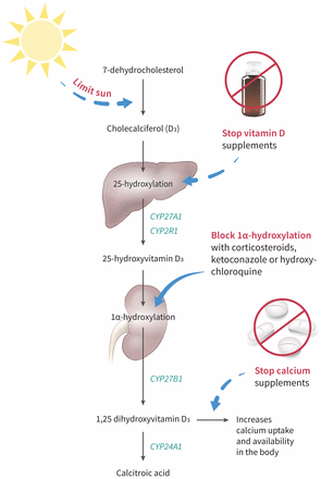
Although vitamin D toxicity is rare owing to a large therapeutic range, its widespread availability in various over-the-counter formulations may pose a substantial risk to uninformed patients. After consumption, vitamin D is carried to the liver where it undergoes hydroxylation and is activated by either microsomal CYP2R1 or mitochondrial CYP27A1 to 25-hydroxyvitamin D3.5,6 The resulting 25-hydroxyvitamin D3 binds to the vitamin D binding protein and is carried to the kidneys for further 1α-hydroxylation by CYP27B1 to produce 1,25 dihydroxyvitamin D3.6 This 1,25 dihydroxyvitamin D3 is transported to target cells and enters the nucleus of the vitamin D receptor, leading to an upregulation in gene expression. Although it is transported by vitamin D binding protein, 1,25 dihydroxyvitamin D3 has a lower affinity to binding relative to 25-hydroxyvitamin D3 and its metabolites.5 A leading hypothesis suggests that an oversaturation of the vitamin D binding protein causes an increase in free active Vitamin D (1,25 dihydroxyvitamin D3), resulting in hypercalcemia.

CYP24A1 plays an important role in the deactivation of 1,25 dihydroxyvitamin D3 to calcitroic acid.6CYP24A1 also breaks down precursor 25-hydroxyvitamin D3 to 24,25-dihydroxyvitamin D3. Loss-of-function mutations in CYP24A1 have been associated with hypercalcemia because of increased vitamin D sensitivity.6 Although we did not perform genetic testing on our patient, it is plausible that he had a CYP24A1 mutation, increasing his susceptibility to vitamin D toxicity.

Manifestations of toxicity

Toxicity can occur over a short period in patients ingesting large doses of vitamin D, either intentionally or inadvertently.7 The literature supports that doses greater than 10 000 IU per day for several months may lead to toxicity (> 200 nmol/L of 25-hydroxyvitamin D3).8 However, differences in patient characteristics such as malabsorption and mutations in CYP24A1 can lead to substantial variation in doses required for toxicity to occur.

Patients may present with symptoms involving the central nervous system and gastrointestinal, genitourinary and cardiovascular systems. Central nervous system manifestations include lethargy, hypotonia, hyporeflexia, confusion and coma. Gastrointestinal symptoms include nausea, vomiting, pancreatitis and constipation; cardiovascular symptoms of toxicity, such as hypertension, arrhythmias and QT segment shortening, may also occur.9 Genitourinary symptoms include polyuria, nephrocalcinosis and renal failure. The symptomatology associated with vitamin D toxicity underscores the suggestion that hypercalcemia may be responsible for most symptoms seen.7Sustained hypercalcemia can also lead to dysregulation of calcium–phosphate homeostasis leading to PTH suppression and impaired bone turnover.3,5

Importantly, patients may be asymptomatic, delaying diagnosis, and abnormalities related to vitamin D toxicity may be detected only incidentally.

In terms of renal involvement, hypercalcemia can cause kidney injury both acutely and chronically. Hypercalcemia can cause acute kidney injury primarily by 2 mechanisms: afferent arteriolar constriction and intravascular volume depletion from a diuretic effect through activation of calcium-sensing receptor at the sodium–chloride cotransporter in the loop of Henle.10 Our patient’s acute kidney injury was likely worsened by volume depletion from diuretic use, prolonged heat exposure and preexisting hypercalcemia from vitamin D toxicity. We saw some improvement in his renal function as his calcium levels decreased, by discontinuing his vitamin D supplements and temporarily holding his diuretics and antihypertensive agents. However, we believe that our patient developed chronic disease as shown by nephrosclerosis on renal biopsy (Figure 1).

Management of toxicity

In cases where vitamin D toxicity is suspected, patients should have their medications — prescribed and over-the-counter — carefully reviewed. Initial strategies to reduce vitamin D levels should focus on reducing dietary or supplementary sources. Although there is currently no evidence from large trials, observational data suggest that clinicians may consider strategies to reduce active vitamin D levels if hypercalcemia persists, through inhibition of 1α-hydroxylase activity (Figure 3).11,12 If patients are asymptomatic, clinicians may choose to monitor levels expectantly, as vitamin D is very fat soluble and levels may take some time to return to normal.
Figure 3:


Several medications have been used successfully to treat hypercalcemia by reducing the active form of vitamin D. Glucocorticoids, ketoconazole and hydroxychloroquine have all been used in cases of hypercalcemia related to sarcoidosis.1,9 Expert opinion suggests that these medications reduce 1α-hydroxylase activity and may be used to manage hypercalcemia by reducing 1,25 dihydroxyvitamin D3 levels.11,12 Given that our patient was reluctant to use glucocorticoid treatment, we used hydroxychloroquine as an alternative to decrease his 1,25 dihydroxyvitamin D3 levels and, in turn, his calcium levels.

Our experience informs us that patients and clinicians should be better informed about the risks regarding the unfettered use of vitamin D. Given new findings from the US Preventive Services Task Force,4 current Canadian guidelines regarding its use in low-risk individuals should be revisited.

Patients with CYP24A1 mutations may be at an increased risk of vitamin D toxicity, and clinicians can consider genetic testing if vitamin D toxicity develops with doses less than 10 000 IU per day. Although vitamin D toxicity is rare, early recognition may prevent chronic complications related to hypercalcemia. In patients who are symptomatic, cessation of supplements along with treatment with glucocorticoids is suggested. In cases where glucocorticoid therapy is not preferred or is contraindicated, ketoconazole or hydroxychloroquine are reasonable alternatives.

The section Cases presents brief case reports that convey clear, practical lessons. Preference is given to common presentations of important rare conditions, and important unusual presentations of common problems. Articles start with a case presentation (500 words maximum), and a discussion of the underlying condition follows (1000 words maximum). Visual elements (e.g., tables of the differential diagnosis, clinical features or diagnostic approach) are encouraged. Consent from patients for publication of their story is a necessity. See information for authors at www.cmaj.ca.

Sunday, April 7, 2019

PKD: Trying At Home Dialysis, Potassium Regulation in PKD, Update: CRISPR Gene Therapy

Living with PKD

From Fox News Channel 5, Atlanta

Kidney failure patient tries at-home dialysis


After 42 years of marriage, Roger and Raynelle Cash of Flowery Branch, Georgia, are not just husband and wife.

They are "care partners," working as a team to give Roger the kidney dialysis keeping the 62-year old alive.

Cash's kidneys failed in 2014, after a 20-year long battle with polycystic kidney disease.

"I was in pretty rough shape when I went into dialysis," he says. "So, even in the dialysis, I started feeling better, because they were taking the excess fluid off me."

At the time, Cash would go to a Fresenius Kidney Care dialysis center 3 days a week at 7 am.

There, he would spend up to 4 hours hooked to a machine filtering the toxins from his blood.

It was grueling.

"You feel pretty drained the days you have dialysis," he says. "And, in-center dialysis has a lot more variation, you feel good, feel real bad, feel good, feel real bad."

After his sessions, Cash would go to his job as a maintenance technician at a manufacturing plant.

But instead of feeling better, he started feeling more rundown.

"The longer I stayed on the in-center, daytime dialysis, the worst I felt," he says. "The weaker I got, the worse I felt."

So, Cash switched to an overnight, or nocturnal, dialysis center, thinking he could sleep through 8-hour sessions.

It didn't work.

"I was not sleeping in the center," he says. "I kept trying to work while I was nocturnal. Ultimately, it basically put me into a physical collapse."

He went on medical leave from his job and grew depressed.

That's when Fresenius Kidney Care offered the Cashes another option.

The company would train Roger and Raynelle to do his dialysis at home, using a machine called a dialyzer.

At first, his nephrologist at the time, Dr. Dinesh Chatoth, who is now the Associate Chief Medical Officer at Fresenius, says Cash was reluctant.

"His concerns were, number one, the machine looks pretty complicated," Chatoth remembers. "And, number two, how am I going to use these needles and stick myself in order to go on dialysis?"

Cash had a painful experience as a young boy when he says a nurse mistakenly gave him a penicillin shot in his arm.

It left him uncomfortable with needles, especially the idea of sticking himself.

But, he decided to try home hemodialysis.

"In all honesty I got desperate," Cash says. "Because I wanted to keep working,"

The Cashes went through 2 and half months of training at Fresenius, learning to set up, clean, and sterilize the machine.

They practiced sticking Roger with the needle.

When the time came for the real thing, Raynelle stuck Roger first, then he took over.

"It turned out to be quite a lot easier than I thought it would be."

Dr. Chatoth says the needles can be challenging for patients.

"There is phobia around that," Chatoth says. "But, once we can break through those barriers, once they experience better health, they tend to thrive and do better on dialysis at home."

Chatoth says Medicare and many insurance providers will pay for at-home dialysis for qualified patients.

He says it's less expensive to do the treatment at home, rather than in a center.

About 12 percent of Frensenius Kidney Care patients do at-home dialysis, Chatoth says.

Most, 10 percent do peritoneal dialysis, through a catheter in their belly.

Only 2 percent use the dialyzer for home hemodialysis, like Cash.

Cash sets his own hemodialysis schedule, 4 nights a week, 3 hours at a time.

He does it after work and before going to bed, in case he feels tired.

"Just about 2 and a half months into it, I woke up and said, 'You know, I feel a lot better,'" Cash says. "And, I felt better days and days in a row."

He and Raynelle have now been using the dialyzer for about 3 years.

Roger Cash has had a few bumps in the road, including a recent bout with colon cancer.

But, he says he's feeling good, and back at work.

"I am not as fast as I used to be, and I'm not as strong as I used to be," Cash says. "Don't have the stamina I used to have, but I can still get the job done. That meant a lot to me."


From Renal and Urology News, by Natasha Persaud

Hyperkalemia Less Likely in PKD Patients


Potassium regulation appears to differ in patients with polycystic kidney disease (PKD) compared with other etiologies of chronic kidney disease (CKD), according to the authors of a new study.

In an analysis of 1788 patients from the KNOW-CKD study (KoreaN cohort study for Outcome in patients With Chronic Kidney Disease), patients with PKD had significantly lower serum potassium levels and lower risks for hyperkalemia than other CKD etiologies during CKD stages 1 to 3b. Hyperkalemia prevalence was also lower among the 293 PKD patients than the other CKD groups up to CKD stage 3 to 4. In adjusted analyses, the risk for hyperkalemia was 5 times higher with diabetic nephropathy, 3 times higher with hypertensive nephrosclerosis, and twice as high with glomerulonephritis, than with PKD.

“Taking these results together, it can be suggested that patients with PKD may have a lower risk of hyperkalemia than those with other etiologies of CKD,” Hyoungnae Kim, MD, of Yonsei University in Seoul, and colleagues stated in BMC Nephrology. They added that further use of dual renin-angiotensin-aldosterone system (RAAS) inhibition may be beneficial in decreasing high intrarenal RAAS activity with a low risk of hyperkalemia in patients with PKD.

The team investigated renal potassium handling by measuring urinary angiotensinogen (AGT), a marker of intrarenal RAAS activity. In multivariable linear regression analysis, having a higher urinary AGT to creatinine (Cr) ratio correlated with lower serum potassium. The ratio appeared significantly higher in PKD patients than in other CKD patients. PKD patients also had a significantly higher transtubular potassium gradient (of urinary to serum potassium) that correlated with the AGT to Cr ratio. In PKD patients, a high urinary AGT to Cr ratio was associated with significantly increased risks for kidney function decline and all-cause mortality by 29%.

“In our study, we also showed that urinary AGT/Cr ratio was correlated with decline in renal function and mortality in patients with PKD. To our knowledge, this is the first longitudinal study that has shown urinary AGT as a prognostic marker in patients with PKD,” Dr Kim and his collaborators stated. The researchers acknowledged that the urinary AGT to Cr ratio can be increased with proteinuria, so additional studies are warranted.


PKD Research

From GEN, Genetic Engineering & Biotechnology News, By MaryAnn Labant

CRISPR Jump-Starts Gene Therapy

Instead of executing extensive overhauls, genomic mechanics are using CRISPR tools to make only the most targeted—and safest—repairs


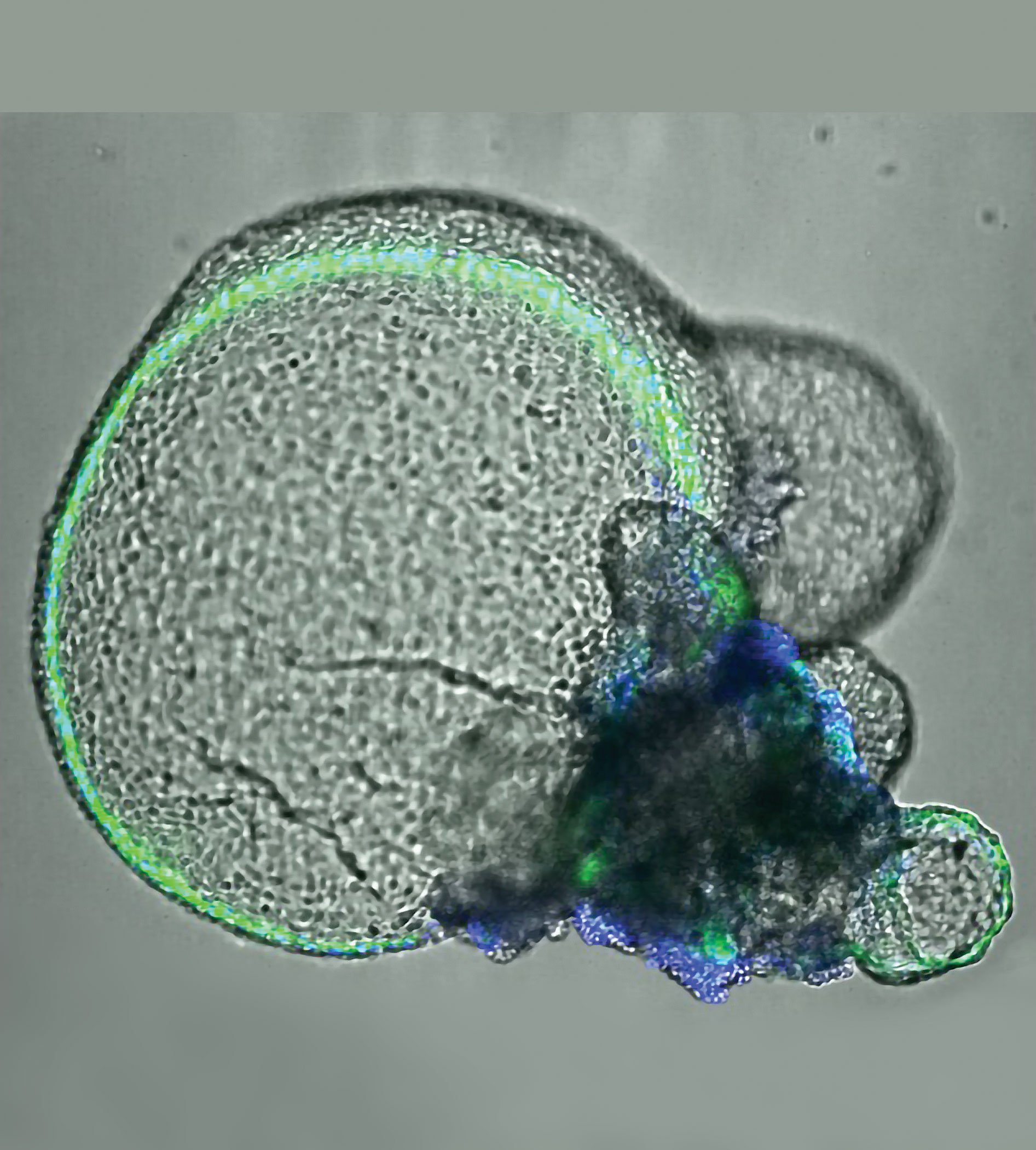
At the University of Washington, Benjamin “Beno” Freeman, PhD, and colleagues are using CRISPR gene editing technology to establish “disease-in-a-dish” models such as this 3D kidney organoid, which shows signs of polycystic kidney disease.


Gene therapy, like a car with mechanical problems, has a history of jerking to life and then quickly stalling. Fortunately, gene therapy has the benefit of a kind of roadside assistance, one that comes in the form of gene editing technology, which is becoming more precise. It can help gene therapy run more smoothly. For example, gene editing can now be used to silence or repair a faulty gene, rather than insert an entire gene into the genome. (If a replacement gene is poorly placed, it can cause insertional mutagenesis.) Several forms of high-precision gene editing can give gene therapy a jump, but the most electrifying form is probably CRISPR.

CRISPR is relatively affordable and easy to use. Consequently, it is being embraced by researchers, who are enthusiastically tinkering with CRISPR system components and developing CRISPR systems that are more efficient—and versatile.

Besides dissecting normal and pathogenic pathways, uncovering biomarkers, and identifying drug targets, CRISPR is starting to translate into the clinic. In February 2019, CRISPR Therapeutics and Vertex Pharmaceuticals achieved a major landmark by announcing the first dosing of a patient with a CRISPR-Cas9 therapeutic in a Phase I/II trial. The therapeutic, CTX001, is being used to treat patients with β-thalassemia. Later this year, the trial will be extended to include patients with sickle-cell anemia.

Other CRISPR biotech companies, notably Editas Medicine and Intellia Therapeutics, are also entering the clinic. These companies, which are focused on gene editing therapies for diseases of the eye and liver, share with Sangamo Therapeutics, Cellectis, and Bluebird Bio an interest in developing earlier forms of gene editing technology. These companies are relying on tools such as zinc finger nucleases (ZFNs), transcription activator-like effector nucleases (TALENs), and meganuclease–TAL effector fusions (megaTALs).

Although the older forms of gene editing technology are still relevant, the relative ease of use and affordability of CRISPR-Cas9 suggests that CRISPR will swiftly prove the most abundant platform for gene therapy. Investigators at Exonics Therapeutics recently reported very promising preclinical data using CRISPR gene editing for treating a canine model of Duchenne muscular dystrophy, in which genetic correction of the dystrophin gene appeared to restore limb function in affected King Charles spaniels, supporting advancement to the clinic.

Not only is CRISPR restarting gene therapy, it is also establishing a virtuous cycle of technological development. CRISPR has advantages that recommend it to a growing user base, which is working to enhance CRISPR strategies, methodologies, and nucleases, which keep broadening CRISPR’s appeal, and so on. This cycle may ultimately bring the benefits of gene therapy to patients, including patients suffering multigene disorders.

Mechanistic insights into kidney disease

The kidney is particularly challenging to study. The early stages of kidney disease are difficult to see, and patients with advanced stages of kidney disease often have secondary complications that affect disease understanding.

“There are many different forms of kidney disease, and there is no magic bullet to cure all of them,” says Benjamin “Beno” Freedman, PhD, assistant professor in the Department of Medicine at the University of Washington. “This is where precision medicine helps us to understand the root causes of disease and to intervene early, before dialysis or a transplant is required. Dialysis is crude compared to a functional organ and typically is not a long-term solution, and there is a lack of transplant organs.”

An experimental system must recapitulate some of the kidney’s complex composition and functions. The Freedman laboratory helped to develop the 3D kidney organoid technology that fulfills this need. Kidney organoids are made from stem cells; multiple cell types develop in a relatively intricate nephron-like arrangement that looks similar to the types of structures seen in the kidneys.

The geometry can differ depending on growth conditions, but all kidney organoids are essentially the same regardless of the laboratory of origin. Results are reproducible from laboratory to laboratory, lending standardization and credibility. Using single-cell RNA sequencing and other tools, researchers have found that organoids naturally make about 15 of the approximately 30 types of cells within the kidney.

“One of the very powerful aspects of CRISPR is that it is a functional tool,” Freedman emphasizes. “It changes an outcome and allows us to manipulate these organoids. This makes it possible to start interrogating and identifying the pathways that are involved in constructing the kidney in a functional way and to recreate disease in the organoids.”

Although there is a contribution of genetics to any form of kidney disease, 10–15% of kidney disease is caused by single-gene mutations. If these mutations could be corrected, a big dent would be made in the number of transplants needed. But CRISPR, while more efficient than other technologies, is still not 100% precise—off-target effects remain a concern to many investigators.

Polycystic kidney disease (PKD) is one of the most common genetic diseases, affecting 1 in 600 people. PKD has few treatments and no cure, making it a good candidate for a gene therapy approach. The genes that cause the disease are known, but it is not known how they normally work in the body.

Using the organoids and CRISPR, the Freedman laboratory has developed a PKD disease-in-a-dish model. The model is shedding light on how PKD occurs at the mechanistic level, leading toward finding interventions to halt disease progression.1

“We have also used CRISPR to produce kidney organoids with fluorescent proteins to better visualize how kidney cells work and the effects of treatments on these cells,” Freedman points out. “CRISPR is an excellent research tool because it allows us to investigate genetic pathways in a way that is far more specific than we were able to do before.”

An approach for monogenic blood disorders

Approximately 75% of all monogenic mutations are caused by point mutations—missense, nonsense, and frameshift mutations. Ideally, these disease genes should be corrected directly at their endogenous loci.

Homology-directed repair (HDR) has low repair efficiency. The dominant repair pathway in cells is usually non-homologous end joining (NHEJ). In theory, this repair mechanism should restore the open reading frame (ORF) that is disrupted by a particular disease mutation in approximately one third of the indels. NHEJ should lead to a significant number of ORF reconstitutions.

“My research results show a gene repair efficiency of up to 25% for some CYBB [cytochrome b-245 beta chain] mutations and an on-target mutation rate of 75% at the endogenous CYBB locus,” says Duran Sürün, PhD, a postdoc in medical systems biology at the Dresden University of Technology. “I am convinced that a donor-template-free, RNA-guided Cas9 endonuclease (RGN) approach has a high potential for personalized gene therapy of chronic granulomatous disease (CGD) and other monogenic blood disorders.”2

Although HDR can be utilized for its precise gene repair mechanism, the efficiency is low and requires a positive selection to enrich for gene-corrected cells. This low efficiency arises from double-strand break (DSB) repair in mammalian cells predominately occurring by NHEJ. More importantly, NHEJ represents the dominant DSB repair pathway in hematopoietic stem and progenitor cells (HSPCs).

Furthermore, the donor template required for HDR introduces a risk of random integration. For that reason, the donor-template-free RGN approach is a better strategy for the potential use in personalized gene therapy, Sürün believes.

Currently, the delivery of Cas9 into stem cells proves to be the greatest challenge. Ideally, RGN delivery for gene therapy should be transient and virus free to avoid insertional mutagenesis and immunological side effects. Special consideration should be given to alternative RGN delivery strategies for future experiments.

Advanced electroporation strategies are most promising. RGNs can be delivered as preassembled gRNA/Cas9 protein complexes. Next steps are to test this strategy in patient-specific HSPCs that will be transplanted after ex vivo gene repair into immunodeficient NSG mice.

Genome-editing tools open a wide range of new possibilities in gene manipulation including target-specific gene repair. The results of the first gene therapy studies in patients will be decisive for the further usage of the CRISPR-Cas9 system. According to Sürün, the new Cas9 protein versions hold great promise because they show less off-target activity in cells.

Merging expression levels with safety

CRISPR-Cas9 is a transformative tool that will continue to operate at the leading edge of basic and translational research. According to Mark J. Osborn, PhD, assistant professor, Department of Pediatrics at the University of Minnesota, a recent study3 had the goal of merging the expression levels achieved with lentiviral transgenesis with the safety of gene editing.

When a viral vector is used, it is common for multiple copies of the vector to deliver their payloads to individual cells. If these payloads include strong transcriptional elements, they can promote high-level, sustained gene expression. These elements, whether they are delivered by retro- or lentiviral vectors, often integrate into the genome with a bias for transcriptionally active regions, causing serious adverse events. In contrast, gene editing can result in precision targeting; however, the endogenous promoter may be comparatively weak, resulting in low-level gene expression.

The Minnesota team inserted a powerful transcriptional element upstream of an endogenous start codon to drive high levels of gene expression with defined integration via HDR. Any nuclease has the potential for off-target effects due to overlapping sequence homology at other genomic loci. To reduce the potential off-target effects, Osborn and colleagues decided to rely on proper targeting to drive expression of a selectable marker that aided in selecting for proper targeting events.

Osborn indicates that his team is further assessing its strategy by defining and mapping potential off-target sites. The hope is that the approach will streamline engineering and make high-level gene expression safer than when integrating vectors are employed.

Identifying off-target effects

Identifying unwanted off-target effects remains an essential requirement for clinical translation of genome editing. Unfortunately, a well-validated method that can reliably identify these events in vivo has been lacking.

Filling this void is a sensitive, unbiased, and generalizable strategy called VIVO (Verification of In Vivo Off-targets). Developed at the Massachusetts General Hospital laboratory of J. Keith Joung, MD, PhD, VIVO allows for the robust identification of genome-wide CRISPR-Cas off-target effects in vivo. VIVO was described in a 2018 Naturearticle,4 which demonstrated that CRISPR-Cas can induce substantial off-target mutations in vivo and that appropriately designed guide RNAs can direct efficient in vivo editing without inducing detectable off-target mutations.

The VIVO approach can be used irrespective of the delivery method for CRISPR-Cas. It is envisioned as a two-step strategy: The first in vitro step uses the CIRCLE-seq method to identify potential off-target cleavage sites of a nuclease of interest on purified genomic DNA.5 Next, off-target sites identified by CIRCLE-seq are examined for evidence of indel mutations in the genomic DNA of target tissues in vivo that have been treated with the nuclease.

The in vivo detection limit of VIVO is limited by the current error rate of sequencing. Previous in vivo studies used computational in silico approaches or the GUIDE-seq method performed on surrogate cells in culture to identify off-target effects.

The results presented by Joung’s group provided the first convincing demonstration that CRISPR-Cas can induce significant off-target mutations in vivo, and, importantly, that these off-target effects can be eliminated with appropriately designed guide RNAs.


Sunday, March 24, 2019

PKD Foundation: Kidney Organoids, Gift of Life: Don't Take Your Organs to Heaven, Medical Marijuana: Illinois, Improving Kidney Care System

PKD Research

From PKD Foundation, by Dr. David Baron, Chief Scientific Officer

PKDF Chief Scientific Officer on kidney organoids: what they are and why they’re important


There are some truly fascinating techniques in the research field being used to help increase our understanding of PKD. This week, our Chief Scientific Officer, Dr. David Baron, shares how the development of kidney organoids may help us get closer to new treatments:

Cell biologists began to grow cells derived from various tissues and organs into what are known as cell cultures in glass dishes (in vitro). These cells could accomplish some of the functions they were designed to do, such as secrete proteins or direct the movement of salts, water, and other across them. However, these cell cultures were mostly two-dimensional and didn’t look anything like the organs or tissues they came from. Another problem, aside from lack of structure, is that these cells would divide only so many times, and with increasing cell division, the cells would often lose their original functions that distinguished them in the first place.


What these biologists really wanted to build was a cell culture system by which the freshly isolated cells could grow into three dimensional structures rather than flat sheets, or even better, grow into structures that, to some extent, reflected where they came from. An early example of this is the use of Madin-Darby Canine Kidney cells or MDCK cells, first established to study viral infections. Scientists later discovered that they could grow these cells into three dimensional tubules that could transport salts and water, functions similar to what the mammalian kidney tubules do. However, these were simple structures of the same cell type that had only a vague resemblance to the complicated kidney from which they were derived.

Dogs are not humans, however. Recent exciting research, in part funded by the PKD Foundation, has shown that human pluripotent stem cells (hPSCs) derived from human skin biopsies can be grown into kidney organoids. Pluripotent stem cells can be induced to change (differentiate) into many distinct cell types. For example, more than 20 distinct cell types required to accomplish the many functions of the human kidney. Much more complex than MDCK cells in culture, these hPSCs can be grown into organoids that resemble in some key ways the structure of the human kidney. The latest research is seeking to grow blood vessels and connective tissue to augment the organoids. Someday this research could be used to actually grow a functional kidney! More importantly for PKD research right now, these organoids can be made to express the ADPKD mutation that causes the disease.


One of the barriers to finding effective treatments for PKD is the lack of an in vitro system to screen potentially promising molecules that could become novel drugs. Such an in vitro system allows for high throughput screening, which is orders of magnitude more efficient than relying on rodent models of PKD. Kidney organoids, particularly those containing the ADPKD mutation, while not quite high throughput, are much more efficient than animal models for screening molecules that could be the next generation of drugs for the treatment of PKD. The PKD Foundation is proud to play a continuing role in this exciting research!


Gift of Life

From Ottawa Citizen, by BLAIR CRAWFORD

'Don't take your organs to Heaven': Orléan's man turns truck into a billboard for organ donors


Three times a week, 52 weeks a year, Steve Dunn makes the trek from his Orléans home and settles into a comfy chair in the basement of the Riverside Hospital. He’ll be there for the next 4 1/2 hours as his blood pumps through a life-saving kidney dialysis machine.

It’s a routine he’s been doing for five years.

“It’s like a part-time job, really,” said Dunn, 47.

Frustrated by his half-decade wait for a kidney donor, Dunn has turned the back of his GMC pickup into a moving billboard for organ donors.

“Asking for help. I need a kidney transplant,” reads the print on the truck tailgate. “Many others like myself need your help. Don’t take your organs to Heaven. Save a life instead.”

Dunn added the slogan to his truck about a month ago, not just to help his own predicament, but for others who are languishing on transplant lists.

“It’s probably not going to help me — although it would be a miracle if it did — but maybe it will help people down the road,” he said. “At least it might make people think about organ donation.”

Dunn is one of 4,300 Canadians waiting for kidney transplant. Kidney transplants are one of the oldest and most common organ transplants. The first successful transplant took place in Illinois in 1950 from a cadaver. Two years later a kidney was successfully transplanted from a living donor.

Today, the operation boasts a success rate of up to 95 per cent, according to the Kidney Foundation of Canada. But there are still not enough organs available.

Dunn suffers from high blood pressure which led to a stroke 10 years ago and eventually to kidney failure. His kidneys now operate at under 10 per cent efficiency, meaning dangerous toxins build up in his blood stream that must be removed through dialysis.

His blood type is O-positive, meaning he can donate blood to anyone, but can only accept blood or an organ from someone else with the same blood type. His sister has diabetes and is ineligible to donate. His mother is too old. His brother is the wrong blood type.

The weekly need for dialysis means the former delivery driver can’t hold a full-time job.

That’s one of the hidden costs that affects so many people with kidney disease, said Elizabeth Myles, executive director of the Kidney Foundation of Canada. Even the cost of travel and parking three times a week at hospital can be prohibitive. People with kidney disease face higher food bills because of the cost of a healthy, kidney-friendly diet that avoids cheaper, processed foods.

“The financial burden of kidney disease is one of the things we want to raise awareness about,” Myles said. “There are a lot of costs passed on to people with kidney disease.”

About four million Canadians, or one in 10, suffer from chronic kidney disease and that number has been increasing, particularly among the young, she said.

For many, like Dunn, a transplant is the best hope for a normal life.

“Even though the vast majority of Canadians support organ donations according to polls, the number who actually consent is relatively small,” Myles said. It’s important that those who do wish to donate organs, register as a donor and let their loved ones know of their wishes.

Dunn is still waiting to hear if a donor is available, but his truck has been turning heads. Once, he came out of a bank at Place d’Orléans and found a man waiting to speak to him.

“He said, ‘I want to give you a kidney’,” Dunn recalled. “He was in a wheelchair but he said that he wanted to help.”

Dunn helped the man pass his name along and though he doubts the man will be a suitable donor, he was touched by the stranger’s selflessness.

“It was unbelievable. You don’t see that very often in the world today.”

In Ontario, organ donation is no longer linked to your driver’s licence. Instead, registration is done through Service Ontario or online at beadonor.ca.

March is Kidney Month in Canada. For more information, visit the Kidney Foundation of Canada at kidney.ca


Living with PKD

From Chicago Tribune, by Robert McCoppin

Proposal would make state medical pot program permanent, add osteoarthritis, chronic pain, autism, migraines, other conditions

Medical marijuana in Illinois would become legal permanently under a proposed new state law, and the list of qualifying conditions would expand greatly to include common conditions such as osteoarthritis, autism and chronic pain.

The former top regulator of medical marijuana in the state, Bob Morgan, is a new Democratic state representative from Deerfield and the chief sponsor of the plan. He says he knows of no significant opposition, and expects lawmakers to approve the measure this spring.

“We’ve had a (pilot program) that shows this is a safe program without diversion (to illegal use),” Morgan said. “It’s helping people who are struggling with serious medical conditions.”

The medical marijuana program allows patients with any of about 40 serious medical conditions, such as cancer and AIDS, to qualify for 2.5 ounces of marijuana every two weeks with their doctors’ approval.

The pilot program has evolved a couple of times since Illinois lawmakers approved it with bipartisan support in 2013, and sales began in late 2015. Former Republican Gov. Bruce Rauner later approved extending the life of the program to mid-2020 and added post-traumatic stress disorder and terminal illness as qualifying conditions, but abolished the medical cannabis advisory board that had recommended adding more conditions.

Last year, Rauner signed a law to allow medical marijuana as an alternative to prescription opioids, and ended a requirement for patient fingerprints and criminal background checks. Those two measures significantly expanded the program to its current size of about 58,000 patients, but so far the program remains smaller than in many other states, mainly because it doesn’t allow patients to use the drug for pain, the most common qualifying condition elsewhere.

The General Assembly is also expected soon to consider legalizing recreational use of cannabis, but if approved it would likely take until next year before sales are allowed. One advantage of medical marijuana would be to avoid taxes placed on recreational marijuana.

By many accounts, the program has worked as intended. Many of the patients say they use marijuana to help them get off other stronger prescription drugs. Police have not reported any major problems with people selling or using medical marijuana without certification.

Even Healthy and Productive Illinois, a nonprofit group which is fighting the proposed legalization of recreational marijuana, does not oppose medical marijuana or decriminalization, lobbyist Tim McAnarney said. He wasn’t able to comment on the proposal to make the medical program permanent, saying he’d have to see how much it would expand use.

The bill would also allow doctors to be paid by medical marijuana businesses or patients. Morgan said that doctors would still be prohibited from getting kickbacks for referrals, but this would allow them to be paid for research.

As proposed, new qualifying conditions would be chronic pain, autism, irritable bowel syndrome, migraines, osteoarthritis, anorexia nervosa, Ehlers-Danlos syndrome, Neuro-Behcet's autoimmune disease, neuropathy, polycystic kidney disease and superior canal dehiscence syndrome.

Research on the effects of cannabis on autism is very limited. Many doctors and several medical groups warn against using a drug which has not been thoroughly tested and approved by the U.S. Food and Drug Administration, and that the federal government classifies as being addictive with no medicinal value.

The American Academy of Pediatrics opposes the use of medical marijuana, noting that it can harm memory, learning and coordination, and lead to psychological problems or drug dependence. But the AAP supports “compassionate use” for children with debilitating or life-limiting diseases, and urges the government to relax the classification of the drug to allow more research.

Mothers Advocating Medical Marijuana for Autism, a nonprofit advocacy group, lists several studies suggesting preliminary support for the treatment, and counting nine states that allow the drug for autism.

Marijuana has been shown to be helpful for many of the conditions that accompany autism, such as epilepsy, pain, aggression and anxiety, said Wendy Fournier, president of the National Association of Autism. She said cannabidiol oil tinctures have helped reduce her autistic daughter’s seizures and helped her sleep.

Recreational marijuana has been legal in Colorado for years. What can Illinois learn before its residents start firing up? »

“There aren’t any good alternatives, so if this is working for some people, go for it, let’s do it,” she said. “This is a potentially life-changing treatment for some people.”

The proposed bill also includes language to allow unspecified “medical conditions or symptoms for which a person may benefit from the use of medical cannabis,” but Morgan said he would remove that language as a compromise to gain broader support.

Those conditions were previously recommended by the former advisory board, and some were the subject of lawsuits and court orders, but the Rauner administration refused to approve them.

Jim Champion, a veteran with multiple sclerosis, was looking forward to changes the bill would bring, such as no longer forcing patients with chronic conditions to recertify their ailments. With his wife Sandy, the Champions lobbied to pass the original law, and worked with Morgan on the new bill.

One important change, they said, was to have the state Department of Public Health make provisions for veterans to qualify under for medical marijuana as an alternative to opioids. Because the drug remains prohibited under federal law, Veterans Affairs doctors are prohibited from certifying their patients.

“We fully support this and look forward to making it permanent,” Sandy Champion said.

The bill would also allow patients to have multiple caregivers who could buy their medical marijuana for them, would allow transportation of the drug after it is opened, and would provide for returns of damaged or inadequate products.

rmccoppin@chicagotribune.com


Kidney Care System

From The Hill, Blog, BY DR. ALLEN R. NISSENSON, OPINION CONTRIBUTOR

Congress, the administration, and kidney care community must further improve care for 30 million Americans


March is National Kidney Month, a perfect time to reflect on the progress made in kidney care and efforts to advance that success to the next level. The challenge facing individuals with kidney diseases requires a united effort from Congress, the Administration, the National Institutes of Health (NIH) and the kidney care community. Fortunately, the collective commitment and momentum are in place, and now is the time to seize on this opportunity to maintain this forward trajectory.

More than a decade ago, the kidney care community set an ambitious goal to improve patient care outcomes and reduce health care costs. We have made significant strides in advancing kidney health with numerous multi-disciplinary work groups consisting of clinicians, providers, patient groups, researchers and manufacturers working together to: develop performance measures for accountability and endorsement through the National Quality Forum process; undertake a successful initiative to improve survival in the first year of dialysis; establish a strategic “quality blueprint” that identifies the leverage points for quality improvement; and publish a framework for considering patient reported outcomes (PROs) in this vulnerable population. All of these initiatives challenged us as a community while we worked constructively with policymakers on meaningful programs and policies that advance us toward the common goal of higher quality care and quality of life for patients with kidney diseases.

Now, a new analysis by Discern Health, which examined data from the government’s United States Renal Data System (USRDS), confirms that patient outcomes have significantly improved during the past decade. All-cause dialysis mortality rates have fallen, as have hospital admission rates for individuals who rely on dialysis care for long-term care needs or while they await a kidney transplant. In fact, gains in survival rates among this population now outpace those for cancer, heart disease, diabetes, and many other serious chronic diseases. Incredibly, the gains in kidney care come even as some chronic diseases saw worsening mortality – heart failure and myocardial infarction among them.

Despite the fact that individuals with kidney failure are among the most complex and costly Medicare beneficiaries to treat, improvements in dialysis care have led to substantial cost savings for Medicare. Discern Health confirmed that Medicare’s spending growth on individuals in kidney failure slowed from 7 percent to 1.5 percent over a 10-year period.These savings persist even after accounting for the fact that the number of Americans who require dialysis has risen sharply in the last few decades, and are now in the billions of dollars.

The kidney care community’s and policymakers’ efforts to advance access, choice, and quality while maintaining the integrity of the Medicare ESRD benefit have had positive, measurable results, yet there is still much more room to improve. We can—and must—do better to build on the real gains we’ve made, and focus on other critical areas such as prevention, access and choice, and increased investment in research and innovations.

In order to challenge ourselves again, we need a bigger and bolder agenda aimed at further boosting survival rates, focusing on patient-centered care and quality of life, improving access to treatment, investing in innovative therapies and medicines, and ensuring kidney health is more effectively addressed before individuals reach kidney failure.

This ambitious agenda requires working collaboratively with policymakers and other stakeholders through public-private partnerships to achieve even greater gains for kidney health. Any constructive collaboration must address not only quality and innovation but also the chronic Medicare underfunding as reported by the independent, nonpartisan Medicare Payment Advisory Commission (MedPAC) in January. This is particularly concerning in the ESRD program as approximately 85 percent of patients rely on government funding for their care.

Last year, the kidney community held a briefing on Capitol Hill with leaders from the kidney care stakeholders, bipartisan members of Congress, and senior representatives from NIH and CMS to share a bold vision for kidney care in America and commit to improving the lives of the tens of millions of Americans living with kidney diseases.

As strong supporters of value in the U.S. health care system, we commend Department of Health and Human Services Secretary Alex Azar’s efforts to foster even greater improvements for the 30 million Americans living with ESRD. Our shared commitment to programs like the Kidney Innovation Accelerator (Kidney X), which is running a competition to develop the new generation of advanced kidney care technology, underscores our commitment to continuously improving kidney health. In addition, we strongly support the interests of HHS and the Center for Medicare & Medicaid Innovation in advancing the care for individuals with chronic kidney diseases before kidney failure occurs, and for significantly increasing the number of patients receiving dialysis at home or a kidney transplant.

The administration has already shown a welcome focus and dedication to prioritize policies and programs to address disease affecting millions of Americans. Secretary Azar vowed in remarks to the National Kidney Foundation that “together, we can deliver much better care, and longer lives, for Americans with kidney disease.” In Congress, a bipartisan group of lawmakers in the House and Senate are working on an updated version of the 2018 Chronic Kidney Disease Improvement in Research & Treatment Act that will be introduced later this year.

As someone who has been a practicing nephrologist for more than 40 years, it is exciting to see the community standing shoulder-to-shoulder with the policy community. We are excited to do our part to advance this bold agenda with a renewed and reimagined commitment to innovation.

To that end, we look forward to continued collaboration with policymakers in Congress and the administration to further help patients with chronic kidney diseases, kidney failure and transplants live longer, healthier, and more fulfilling lives.

Nissenson is chair of Kidney Care Partners.

Sunday, March 10, 2019

Kidney Health: Check Your Urine Color, PKD Awareness Month: Planters and Art, 3D Printed Kidney Proximal Tubules

Kidney Health

From Financial Review, by Hilary Brueck


Whether you're trying a keto plan, a new plant-based routine, intermittent fasting, or the Whole30, experts agree: dieting is hard work.

Thomas Chi, a doctor at the University of California, San Francisco, urology department, said proper hydration tends to be one of the toughest things for his dieting patients to remember when they start a new weight-loss plan.

"People who are on extreme diets, they tend to have higher levels of salt and less hydration," Chi said.

Fortunately, your body is here to help. All you have to do is take a little time to listen to the brain's subtle cues: If you're feeling thirsty, drink some water.

This is especially important if you're eating more meats and high-salt foods, Chi said, because a combination of "tons and tons of meat" and not enough water could lead to kidney stones, especially if you have a family history of the issue.

"I'm not seeing this enormous uptick of patients with kidney stones because they're all doing a keto diet now," he said.

But he still thinks it's a good idea to pay attention to when your body's asking for water.

No matter if you're on an extreme diet or no diet at all, that's generally good advice.

"A lot of times, people just seem to be a little dehydrated," the dietitian Jason Ewoldt from the Mayo Clinic previously told Business Insider.

But the popular notion that you must down eight full glasses of water a day is bogus. And nobody really needs any special sports drinks, even after a workout.

Your body knows exactly when you need more water

Just drink when you're thirsty because the body is designed to know precisely when you need more water.

There's actually a "thirst centre" in the brain that helps regulate thirst, and it's impressively tuned.

Even though it takes tens of minutes for the body to hydrate once cool water touches your lips, drinking quenches thirst within seconds.

The brain's thirst cues can be relatively mild and easily mistaken for hunger pangs, or just ignored. Pretty soon, that can prompt dehydration and lead to overeating.

A 2016 study of more than 18,000 people in the US found that those who drank more water were consistently more satisfied and ate fewer calories daily. (Drinking with a meal both helps replenish saliva and aids digestion.)

Steady water drinkers also consumed lower amounts of sugar, fat, salt and cholesterol than dehydrated people.

So if you're unsure if you're feeling peckish, try sipping on some water, waiting about 15 minutes, and checking if your stomach really is rumbling.

"If you were truly hungry, you might still feel a stomach pang, whereas if you were just thirsty, you'll feel satisfied," the Polycystic Kidney Disease Foundation said.

If you're not a big fan of water, you can dress up your drink by adding lemon juice into your glass.

That's a cheap, doctor-recommended hack to help prevent kidney-stone formation.

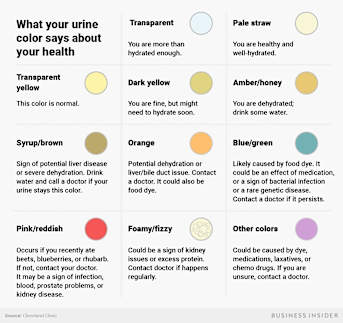
No need to fuss too much over the colour of your urine, either. As long as it's not a super-dark yellow or an amber colour, you're doing well.

This story first appeared in Business Insider. Read it here or followBusinessInsider Australia on Facebook.

BusinessInsider.com.au


From Science Daily

Growing evidence: Water as a potential treatment for inherited cause of kidney failure


People with polycystic kidney disease (PKD) could benefit from a moderate increase in water intake, according to new research.


A study from The Westmead Institute for Medical Research found that a moderate increase in water intake in rats with PKD led to a long-term reduction in kidney cyst growth and fibrosis.

This latest findings add to the growing body of evidence that supports water as a safe and effective treatment for PKD.

Polycystic kidney disease is the most common inherited cause of end-stage kidney disease. It is a chronic condition, in which fluid-filled cysts damage healthy tissue and kidney function.

Left untreated, it can cause complications, including high blood pressure, heart problems and, in severe cases, kidney failure.

More than 2,000 Australians with PKD currently receive dialysis or need a kidney transplant.

Lead researcher Dr Priyanka Sagar said that water may be a potential treatment for PKD, because it stops the hormone responsible for cyst growth.

"Previous studies in animals haven't shown whether this benefit continues over time, and there is presently no evidence in humans," Dr Sagar said.

"Our research in rats showed that increased water intake reduces the long-term progression of cyst growth and kidney fibrosis when administered during the early stages of kidney disease.

"Significantly, we identified that only a moderate increase in water was needed to have this sustained benefit in rats."

The research also showed that increased water intake had secondary benefits for some complications associated with PKD.

"Interestingly, we found that increased water intake also reduced hypertension," Dr Sagar said.

"PKD is linked to an increased risk of cardiovascular disease, so this is an important protective effect."

Currently, treatment options for PKD in humans are limited. Dr Sagar said that further studies are needed in humans to prove that water is an effective treatment for kidney cysts.

"We're finding more evidence to support water as a viable treatment for PKD," she said.

"However, further studies are needed to determine its effectiveness.

"Water is cheap and accessible, so the idea that it could be used as a treatment for PKD in the future is very exciting," she concluded.


Story Source:

Materials provided by Westmead Institute for Medical Research. Note: Content may be edited for style and length.


From MD Linx

Long-term trajectory of kidney function in autosomal-dominant polycystic kidney disease

In a 14-year observational cohort study (The Consortium for Radiologic Imaging Studies of Polycystic Kidney Disease study), researchers assessed the trajectories of glomerular filtration rate (GFR) decline in 241 adults with autosomal dominant polycystic kidney disease (ADPKD). Taking into account baseline age and magnetic resonance imaging measurements of total kidney volume (TKV) according to the method of Irazabal, 5 subclasses of participants were defined, as an estimate of the rate of kidney growth. Using mixed polynomial models, they reconstructed and fitted GFR trajectories spanning over four decades of life. For validation, they used data from the HALT-PKD study. Findings revealed a nonlinear GFR decline in ADPKD. A single measurement of kidney volume could predict GFR decline trajectory throughout adulthood. The possible utility of these models was suggested for clinical prognostication, clinical trial design, and patient selection for clinical interventions. Growth in kidney volume was causally related to GFR decline. The use of TKV as a surrogate endpoint in clinical trials was supported.


Read the full article on Kidney International


PKD Research

From ECN Magazine, by Harvard University


Immunofluorescence staining of a 3D bioprinted vascularized proximal tubule with a proximal tubule epithelial marker stained in green in the proximal tubule channel and a vascular endothelial marker stained in red in the adjacent vascular channel. The magnified cross-section illustrates that the two different cell types form luminal perfusable structures in their respective channels. Credit: Wyss Institute at Harvard University


Every day our kidneys tackle the daunting task of continuously cleaning our blood to prevent waste, salt and excess fluid from building up inside our bodies. To achieve this, the kidneys' approximately one million filtration units (glomeruli) first remove both waste products and precious nutrients from the blood stream, then specialized structures known as the proximal tubules reabsorb the "good" molecules returning them to our bloodstream. While the reabsorptive functions of the proximal tubule can be compromised by drugs, chemicals, or genetic and blood-borne diseases, our understanding of how these effects occur is still limited.

To enable the study of renal reabsorption outside the human body, Wyss Institute Core Faculty member Jennifer Lewis, Sc.D., and her team working within the Wyss Institute's 3D Organ Engineering Initiative that she co-leads, and in collaboration with the Roche Innovation Center Basel in Switzerland, created a 3D vascularized proximal tubule model in which independently perfusable tubules and blood vessels are printed adjacent to one another within an engineered extracellular matrix. This work builds upon a continuously perfused 3D proximal tubule model reported earlier by the team that still was lacking a functional blood vessel compartment. Using their next-generation device, the team has measured the transport of glucose from the proximal tubule to the blood vessels, along with the effects of hyperglycemia, a condition associated with diabetes in patients. Their study is published in the Proceedings of the National Academy of Sciences (PNAS).

Lewis is also the Hansjörg Wyss Professor of Biologically Inspired Engineering at Harvard John A. Paulson School of Engineering and Applied Science (SEAS), the Jianmin Yu Professor of Arts and Sciences and a member of the Harvard Stem Cell Institute.

"We construct these living renal devices in a few days and they can remain stable and functional for months," said first-author Neil Lin, Ph.D., who is a Roche Fellow and Postdoctoral Fellow on Lewis' team. "Importantly, these 3D vascularized proximal tubules exhibit the desired epithelial and endothelial cell morphologies and luminal architectures, as well as the expression and correct localization of key structural and transport proteins, and factors that allow the tubular and vascular compartments to communicate with each other."

As a first step towards testing drugs and modeling diseases, the team induced "hyperglycemia", a high-glucose condition typical of diabetes and a known risk factor for vascular disease, in their model by circulating a four-fold higher than normal glucose concentration through the proximal tubule compartment. "We found that high levels of glucose transported to endothelial cells in the vascular compartment caused cell damage," said Kimberly Homan, Ph.D., a co-author on the study and Research Associate in Lewis' group at the Wyss Institute and SEAS. "By circulating a drug through the tubule that specifically inhibits a major glucose transporter in proximal tubule epithelial cells, we prevented those harmful changes from happening to the endothelial cells in the adjacent vessels."

The team's immediate focus is to further scale up these models for use in pharmaceutical applications. "Our system could enable the screening of focused drug libraries for renal toxicity and thus help reduce animal experiments," said Annie Moisan, Ph.D., a co-author and industry collaborator on the study, and Principal Scientist at Roche Innovation Center Basel. "I am thrilled by the continued efforts from us and others to increase the physiological relevance of such models, for example by incorporating patient-specific and diseased cells, since personalized efficacy and safety are the ultimate goals of predicting clinical responses to drugs."

"Our new 3D kidney model is an exciting advance as it more fully recapitulates the proximal tubule segments found in native kidney tissue," said Lewis. "Beyond its immediate applications for drug screening and disease modelling, we are also exploring whether these living devices can be used to augment kidney dialysis." Currently, life-saving dialysis machines filter blood, but they are unable to retrieve precious nutrients and other species from the filtrate that the body needs for many of its functions, which can cause specific deficiencies and complications down the line. Lewis and her colleagues believe that 3D bioprinted vascularized tubules may lead to improved renal replacement therapies.

"This study presents a significant step forward in human kidney engineering that enables human disease and drug-related studies to be carried out over extended periods of time in vitro. It also represents a major step forward for the Wyss Institute's 3D Organ Engineering Initiative, which aims to generate functional organ replacements with enhanced functionalities for patients in need," said Wyss Institute Founding Director Donald Ingber, M.D., Ph.D., who is also the Judah Folkman Professor of Vascular Biology at HMS and the Vascular Biology Program at Boston Children's Hospital, as well as Professor of Bioengineering at SEAS.


PKD Awareness

From Sylvan Lake News, Alberta, Canada, by KAYLYN WHIBBS
Sylvan Lake’s Winter Village planter contest helps spread awareness


March is Polycystic Kidney Disease Awareness month


The winner of Best Overall Non-Profit in the planter contest with the 2018 Waterfront Winter Village has a very personal touch behind its tree.

Sylvan Lakes’ Rosalie Thon, and her friends, Evan Masse, Trish Jenks and her son Clayton William, designed the planter for kidney disease and transplant awareness.

Thon has Polycystic Kidney Disease and spent four years doing dialysis while waiting to get a new kidney.

She did a battery of tests and had four calls before her kidney came Sept. 21, 2017.

“In that part of my journey I met Evan [Masse], he was my roommate [in the hospital] and he as well had a kidney transplant, and so we just became really good friends and when the planter came up I just thought I wanted to give something back,” said Thon in an interview, adding she has learned a lot throughout her journey.

Masse lives north of Edmonton, but made the trip down to help decorate the tree.

“I know for a fact a lot of people don’t know a lot about kidney disease and/or transplant, so I thought what a better opportunity than to take advantage of this tree,” explained Thon.

The blue and green colouring of the tree was inspired by the colours of The Kidney Foundation of Canada and was decorated with kidney shaped stress balls and cards with ribbon pins for awareness.

“Rosalie and I decided to give people awareness of donating organs, it is a lifesaver for people even though it is sad that some people have to sacrifice their life to save other people,” said Masse in a letter, adding he is thankful for all the people who donate.

Decorating the tree was a two-day project and represented transplant awareness right down to its roots after they removed the original little tree to transplant in an artificial one.

The Kidney Foundation of Canada, Northern Alberta and The Territories branch in Edmonton donated the stress kidneys and cards to use as decorations.

This was the group’s first time entering a planter in the competition and is very proud to have been voted number one in their category.

Thon says when she sets out to do something she always wants to be a high achiever, so she told the group she wanted to win.

“I wanted to win not out of arrogance, but just to get the story out even more,” said Thon, adding she also likes being creative.

Thon and Masse used the lighting ceremony and the planter along the lakeshore throughout the winter as a talking point to help raise awareness, knowledge and share their story.

She said it was exciting to be able to make connections with people and spread the word about kidney disease and transplant.

Since polycystic is hereditary her son, Clayton William, also has polycystic kidneys.

Thon explained hers started with a 2.5 cm cyst, which took 20 years to almost completely shut down her kidney function, so it is hard to tell when William will need to go through the dialysis and transplant procedure.


From The Daily Bruin, UCLA

Art exhibit sheds light on lives of those affected by genetic disorders

The exhibit was created to provide more insight into the lives of patients affected by genetic disorders. (Lauren Man/Daily Bruin)


An art exhibit in Powell Library showcases the hidden impact genetic disorders often have on the lives of UCLA students.

The exhibit, which opened Feb. 19 and will remain open through March in the Powell Library rotunda, showcases various forms of art submitted by five students who have genetic disorders such as lymphoma and cystic fibrosis.

Rushna Raza, a fourth-year molecular, cell, and developmental biology student, said she created the exhibit because of her interest in genetics and because she wanted to provide more insight into the lives of patients affected by these disorders.

“I believe the genetic and the social perspectives are both important because patients’ lives go beyond what meets the eye,” she said. “Those stories never get told unless you ask.”

The exhibit features five pieces of visual and interactive artwork created by students that aim to educate others on the invisible suffering caused by genetic disorders, Raza said. Some students chose to focus on illnesses that affect them directly, while others explored genetic diseases that have affected their loved ones. The projects also reflected the roles these heritable diseases have played throughout their families’ histories.

Chelsea Krob, a fourth-year art student who has autosomal dominant polycystic kidney disease, created a multimedia gallery to raise awareness of her illness.

Her project, “Through Your Journey,” tells the story of her struggle with PKD through photos and a digital recording of a conversation between Krob and her father, who died from the same illness in May.

“My project aims to look at the intergenerational effects of this disease and also my unique relationship with my dad, since we both have (PKD),” she said.

Krob’s portion of the exhibit also features a wall on which observers can add their handprints with ink. She said she was inspired by a previous project concept in which she planned to represent every day of her father’s life through thumbprints.

Raza said the handprint wall represents how everyone is unique due to small differences in their genomes. Similarly, even though human handprints have the same basic shape, small variations make them unique. Raza said this aspect of the exhibit allows students to participate in and leave their own mark on the artwork.

Krob said she hopes observers can gain a better understanding of the lives of people with PKD through her project.

“I used multiple approaches to give the viewer, wherever they’re at with life or death, an entry point into the work,” she said.

Hollie St. Claire, who graduated from UCLA in fall 2018, portrayed the impact of cystic fibrosis in her life through her carving, “Breaking Through.” Three of St. Claire’s siblings have the disease and she recently learned she is a carrier. She said she hopes to show that people with cystic fibrosis are strong and that there is hope for a cure.

To demonstrate the strength of those with cystic fibrosis, St. Claire created a painted carving that depicts an individual pushing against an arch containing strands of DNA. She said she aims to challenge the notion that individuals with cystic fibrosis are weak or fragile.

“The individual in my carving is breaking through the barriers of genetic restrictions,” she said.

Ketana Chadalavada, a fourth-year psychology student who designed the exhibit’s online graphics, said her painting “Survivor” represents her struggle with lymphoma, which she was diagnosed with last year.

She said her portrayal of a bald figure demonstrates her reclaiming ownership of her body. The lime green ribbon over the figure’s heart is an official symbol of lymphoma awareness and ties the painting to the broader lymphoma community.

“I really wanted to show the mental side to my story, including how I coped with (lymphoma) and what I learned from it,” Chadalavada said.

In addition to raising awareness for genetic disorders, Raza added she created the exhibit because she was interested in combining science and art. She said even though she does not consider herself to be an artist, she is proud to have put together a project that allows students to tell their personal stories through art.

“I wanted this project to be a chance for students to share that intimate part of their history through an artistic platform,” she said.