From Scicast.com
Study Shows How and Why Hairlike Structures on Cells are Lost
Many of our cells are equipped with a hairlike "antenna" that relays information about the external environment to the cell, and scientists have already discovered that the appearance and disappearance of these so-called primary cilia are synchronized with the process of cellular duplication, called mitosis. Now, cell biologists at Johns Hopkins report the discovery of new information about how this "hair loss" and cell duplication are linked through the dramatic clipping of the tips of the cilia -- what the scientists dub decapitation -- that begins their disassembly.
The researchers say the new information is key to better understanding how cells decide to undergo mitosis, a process integral to the development of organisms, the maintenance of tissues and the formation of cancer. They also hope their work will shed light on cilia-related diseases, like polycystic kidney disease, and certain forms of intellectual disability.
"The decapitation of cilia had been observed before but never explored," says Dr. Takanari Inoue, associate professor of cell biology at the Johns Hopkins University School of Medicine, who led the study. "We now know that it's a normal process, not just something that happens under certain experimental conditions. And we have identified the molecular players that drive it."
A summary of his team's findings appears Jan. 12 in the journal Cell.
Like antennas on minivans, primary cilia are tiny structures that appear on cells within the kidney, brain, retina and inner ear, among other organs, and provide cells with information about the flow and chemistry of fluids outside.
When a cell is not dividing, a state called quiescence, most cellular structures are continuously changing, but primary cilia are relatively stable. Inoue and his group sought to understand what causes the "haircut" when a cell exits quiescence and begins mitosis, during which replicated chromosomes divide and populate two new "daughter" cells.
They started by comparing cells with more and less of an enzyme called Inpp5e, which previous studies had implicated in the stability of cilia. After adding a fluorescent tag to the membrane of cilia in quiescent mouse embryonic cells, the team videotaped cilia's tips. They were seemingly being severed and drifting away once every several hours.
The rate of decapitation was higher in cells missing the Inpp5e gene and when cells were given signals that stimulate mitosis. But when the scientists added a molecular code to the Inpp5e protein so that it would collect in the cilia, the rate of decapitation greatly slowed. Inoue says this suggests that the presence of Inpp5e in the cilia stalls the decapitation process and that mitotic signals expel Inpp5e from the cilia to promote decapitation.
"Because genetic mutations in Inpp5e are associated with Joubert syndrome, which is characterized by abnormal brain development and intellectual disability, we now suspect that Inpp5e affects brain development," says Inoue.
The function of Inpp5e is to deplete a fatlike molecule, called PIP2, from the membrane of cilia. A fluorescent tag that binds to PIP2 showed the researchers that the concentration of PIP2 in the cilia, especially near the tip, was synchronized too: PIP2 accumulated at the tips of cilia after cells received mitotic signals, and the cilia were clipped off at the site of PIP2 accumulation.
Because PIP2 plays a key role in the formation of wirelike structures made from the protein actin, the team used a fluorescent biosensor to measure and view the structures' formation in cilia. In cilia containing excess PIP2 due to a lack of Inpp5e, the researchers found a tenfold increase in wire formation. In addition, they saw that the wire formation happened right at the site of decapitation just a few minutes ahead of time.
"We think that wire formation is actually providing the force that decapitates the cilia," says Inoue.
To see how decapitation affected the rest of cilia disassembly, Siew Cheng Phua, a graduate student in Inoue's lab and the primary author of the paper, designed a way to stop wires from forming within cilia. She found that the wires are essential to cilia decapitation and that decapitation is required for the full disassembly of cilia.
Next, the team analyzed the timing of cilia decapitation. Using a rainbow of fluorescent tags, the researchers followed cells with cilia from quiescence through their transition to mitosis. The cilia were marked in blue, and the nuclei of the cells changed from yellow to red to dark as they progressed. In normal cells, cilia decapitation generally occurred while cells were still in the quiescent state (yellow nuclei), before transitioning into mitosis (dark nuclei). But in cells whose cilia were prevented from forming wires, the inability to decapitate cilia was associated with a slower transition from quiescence to mitosis.
"Somehow, decapitation triggers the shrinking of the cilia; they shrink by more than what is clipped off," says Inoue. "At the same time, that seems to signal to the cell that it's time to exit quiescence and begin division."
When they analyzed the contents of the decapitated cilia tips -- a collaborative project with researchers in Japan -- they mostly found molecules important for cell signalling and cilia growth, implying that decapitation adjusts the composition and function of cilia.
The clipped tips of cilia might also have clinical implications, according to Phua. Recent research shows that urine from patients with polycystic kidney disease contains tiny sacs enclosed in membrane. Inoue says it's possible they are cilia tips and that they play a role in disease development, but more research is needed to confirm this idea.
Phua adds that: "Abnormal sensory functions of cilia are associated with some cancers, like skin and brain cancer. The novel link between cilia decapitation and mitosis could help us understand how defects in primary cilia influence the abnormal cell divisions which underlie cancer formation.”
Article adapted from a Johns Hopkins Medicine news release.
Living with PKD
From NIDDK (National Institute of Diabetes and Digestive and Kidney Diseases) of U.S. Department of Health and Human Services
Acquired Cystic Kidney Disease
What is acquired cystic kidney disease?
Acquired cystic kidney disease happens when a person's kidneys develop fluid-filled sacs, called cysts, over time. Acquired cystic kidney disease is not the same as polycystic kidney disease (PKD), another disease that causes the kidneys to develop multiple cysts.
Acquired cystic kidney disease occurs in children and adults who have
chronic kidney disease (CKD)—a condition that develops over many years and may lead to end-stage kidney disease, or ESRD. The kidneys of people with CKD gradually lose their ability to filter wastes, extra salt, and fluid from the blood properly.
end-stage kidney disease—total and permanent kidney failure that requires a kidney transplant or blood-filtering treatments called dialysis.
The cysts are more likely to develop in people who are on kidney dialysis. The chance of developing acquired cystic kidney disease increases with the number of years a person is on dialysis. However, the cysts are caused by CKD or kidney failure, not dialysis treatments.
More information is provided in the NIDDK health topics, kidney failure and dialysis.
What are the differences between acquired cystic kidney disease and polycystic kidney disease?
Acquired cystic kidney disease differs from PKD in several ways. Unlike acquired cystic kidney disease, PKD is a genetic, or inherited, disorder that can cause complications such as high blood pressure and problems with blood vessels in the brain and heart.
The following chart lists the differences:
People with Polycystic Kidney Disease
are born with a gene that causes the disease
have enlarged kidneys
develop cysts in the liver and other parts of the body
People with Acquired Cystic Kidney Disease
do not have a disease-causing gene
have kidneys that are normal-sized or smaller
do not form cysts in other parts of the body
In addition, for people with PKD, the presence of cysts marks the onset of their disease, while people with acquired cystic kidney disease already have CKD when they develop cysts.
More information is provided in the NIDDK health topic, Polycystic Kidney Disease.
How common is acquired cystic kidney disease?
Acquired cystic kidney disease becomes more common the longer a person has CKD.
About 7 to 22 percent of people with CKD already have acquired cystic kidney disease before starting dialysis treatments.
Almost 60 percent of people on dialysis for 2 to 4 years develop acquired cystic kidney disease.1
About 90 percent of people on dialysis for 8 years develop acquired cystic kidney disease.1
What causes acquired cystic kidney disease?
Researchers do not fully understand what causes cysts to grow in the kidneys of people with CKD. The fact that these cysts occur only in the kidneys and not in other parts of the body, as in PKD, indicates that the processes that lead to cyst formation take place primarily inside the kidneys.
What are the signs and symptoms of acquired cystic kidney disease?
A person with acquired cystic kidney disease often has no symptoms. However, the complications of acquired cystic kidney disease can have signs and symptoms.
What are the complications of acquired cystic kidney disease?
People with acquired cystic kidney disease may develop the following complications:
an infected cyst, which can cause fever and back pain.
blood in the urine, which can signal that a cyst in the kidney is bleeding.
tumors in the kidneys. People with acquired cystic kidney disease are more likely than people in the general population to have cancerous kidney tumors. However, the chance of cancer spreading is lower in people with acquired cystic kidney disease than that of other kidney cancers not associated with acquired cystic kidney disease, and the long-term outlook is better.
How is acquired cystic kidney disease diagnosed?
A health care provider may diagnose a person with acquired cystic kidney disease based on
medical history
imaging tests
Medical History
Taking a medical history may help a health care provider diagnose acquired cystic kidney disease. A health care provider may suspect acquired cystic kidney disease if a person who has been on dialysis for several years develops symptoms such as fever, back pain, or blood in the urine.
Imaging Tests
To confirm the diagnosis, the health care provider may order one or more imaging tests. A radiologist—a doctor who specializes in medical imaging—interprets the images from these tests, and the patient does not need anesthesia.
Ultrasound uses a device, called a transducer, that bounces safe, painless sound waves off organs to create an image of their structure. A specially trained technician performs the procedure in a health care provider's office, an outpatient center, or a hospital. The images can show cysts in the kidneys as well as the kidneys' size and shape.
Computerized tomography (CT) scans use a combination of x-rays and computer technology to create images. For a CT scan, a nurse or technician may give the patient a solution to drink and an injection of a special dye, called contrast medium. CT scans require the patient to lie on a table that slides into a tunnel-shaped device where an x-ray technician takes the x-rays. An x-ray technician performs the procedure in an outpatient center or a hospital. CT scans can show cysts and tumors in the kidneys.
Magnetic resonance imaging (MRI) is a test that takes pictures of the body's internal organs and soft tissues without using x-rays. A specially trained technician performs the procedure in an outpatient center or a hospital. Although the patient does not need anesthesia, a health care provider may give people with a fear of confined spaces light sedation, taken by mouth. An MRI may include the injection of contrast medium. With most MRI machines, the patient will lie on a table that slides into a tunnel-shaped device that may be open-ended or closed at one end. Some machines allow the patient to lie in a more open space. During an MRI, the patient, although usually awake, must remain perfectly still while the technician takes the images, which usually takes only a few minutes. The technician will take a sequence of images from different angles to create a detailed picture of the kidneys. During the test, the patient will hear loud mechanical knocking and humming noises from the machine.
Sometimes a health care provider may discover acquired cystic kidney disease during an imaging exam for another condition. Images of the kidneys may help the health care provider distinguish acquired cystic kidney disease from PKD.
How is acquired cystic kidney disease treated?
If acquired cystic kidney disease is not causing complications, a person does not need treatment. A health care provider will treat infections with antibiotics—medications that kill bacteria. If large cysts are causing pain, a health care provider may drain the cyst using a long needle inserted into the cyst through the skin.
When a surgeon transplants a new kidney into a patient's body to treat kidney failure, acquired cystic kidney disease in the damaged kidneys, which usually remain in place after a transplant, often disappears.
A surgeon may perform an operation to remove tumors or suspected tumors. In rare cases, a surgeon performs an operation to stop cysts from bleeding.
Have Regular Screenings to Look for Cyst or Tumor Growth
Some health care providers recommend all people with end-stage kidney disease get screened for kidney cancer using CT scans or MRIs after 3 years of dialysis. People with acquired cystic kidney disease should talk with their health care provider about when to begin screening.
Eating, Diet, and Nutrition
No specific diet will prevent or delay acquired cystic kidney disease. In general, a diet designed for people on hemodialysis or peritoneal dialysis reduces the amount of wastes that accumulate in the body between dialysis sessions.
More information is provided in the NIDDK health topics, Eat Right to Feel Right on Hemodialysis and Nutrition for Advanced Chronic Kidney Disease in Adults.
Points to Remember
Acquired cystic kidney disease happens when a person's kidneys develop fluid-filled sacs, called cysts, over time.
Acquired cystic kidney disease occurs in children and adults who have
chronic kidney disease (CKD)
end-stage kidney disease (ESRD)
People with acquired cystic kidney disease may develop the following complications:
an infected cyst, which can cause fever and back pain
blood in the urine, which can signal that a cyst in the kidney is bleeding
tumors in the kidneys
To confirm the diagnosis, the health care provider may order one or more imaging tests:
Ultrasound
Computerized tomography (CT) scan
Magnetic resonance imaging (MRI)
If acquired cystic kidney disease is not causing complications, a person does not need treatment.
A health care provider will treat infections with antibiotics—medications that kill bacteria.
If large cysts are causing pain, a health care provider may drain the cyst using a long needle inserted into the cyst through the skin.
A surgeon may perform an operation to remove tumors or suspected tumors. In rare cases, a surgeon performs an operation to stop cysts from bleeding.
References
[1] Torres VE, Grantham JJ. Cystic diseases of the kidney. In: Taal MW, ed. Brenner & Rector's The Kidney. 9th ed. Philadelphia: W.B. Saunders; 2011: 1626–1667.
[2] Chapman AB, Rahbari-Ouskoui FF, Bennett WM. Acquired cystic disease of the kidneys in adults. UpToDate website. www.uptodate.com . Updated January 14, 2014. Accessed August 8, 2014.
Clinical Trials
The National Institute of Diabetes and Digestive and Kidney Diseases (NIDDK) and other components of the National Institutes of Health (NIH) conduct and support research into many diseases and conditions.
What are clinical trials, and are they right for you?
Clinical trials are part of clinical research and at the heart of all medical advances. Clinical trials look at new ways to prevent, detect, or treat disease. Researchers also use clinical trials to look at other aspects of care, such as improving the quality of life for people with chronic illnesses. Find out if clinical trials are right for you .
What clinical trials are open?
Clinical trials that are currently open and are recruiting can be viewed at www.ClinicalTrials.gov
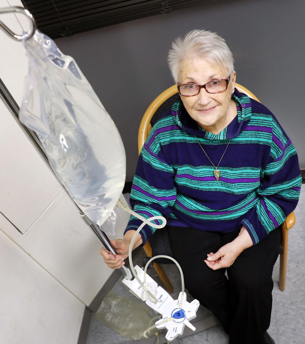
JoAnne Jeffs, 71, started receiving hemodialysis in July 2011. This treatment for kidney failure, which uses a machine to send a patient's blood through a filter to clean it, left the Hinton, Iowa, woman feeling weak, tired and dehydrated. She didn't think she'd make it to Christmas that year.
"I slept a lot," said Jeffs, who has polycystic kidney disease, a condition which causes clusters of cysts to develop in the kidneys.
At age 60, Jeffs' body began to shut down. Her skin took on a gray tone and she lost her appetite. The diagnosis was kidney failure.
A prior bout with breast cancer took Jeffs out of the running for a kidney transplant. She partially regained her kidney function only to begin hemodialysis a couple years later.
Today, Jeffs isn't sure she would even want a donor kidney. She said switching from hemodialysis to peritoneal dialysis gave her back her life. This treatment, which can be done in the comfort of a kidney patient's home, has grown in popularity over the last decade.
According to the U.S. Renal Data System, peritoneal dialysis is less expensive than hemodialysis. One year of hemodialysis can cost up to $72,000, while a year of peritoneal dialysis costs about $53,000.
Renal Associates will begin offering a home peritoneal dialysis training program for patients and their loved ones in March in Dakota Dunes. Learning the process takes about six weeks.
"We are going to be pushing toward this modality because of the fact that we feel that it is going to be better for our patients in the long run," said Ashar Luqman, Jeffs' nephrologist.
"Patients will be much more well-versed in dealing with all the problems which come with dialysis if they are doing it themselves."
The National Kidney Foundation estimates that over 200,000 people use dialysis on an ongoing basis.
Luqman said a young patient recently diagnosed with kidney failure due to polycystic kidney disease, lupus or high blood pressure could survive for years on dialysis if the patient has no other medical problems. He said many patients who are afraid of dialysis say they would rather die than undergo the rigorous treatment.
"I cannot tell you how many times I've had patients when they initially see me they say, 'Absolutely no dialysis,'" Luqman said. "But when they start getting sick and the kidneys shut down, at that time it's too little, too late because they've already gotten to a point where even if we start doing dialysis they're not going to feel a lot better."
Two types of dialysis
Any form of dialysis is better than no dialysis, according to Jeffs. While hemodialysis didn't work well for her, she acknowledged that some people have success with it.
Patients undergoing hemodialysis visit a hospital or dialysis center three days a week. They sit in a chair for 4 1/2 hours as a filter or dialyzer used in conjunction with a dialysis machine removes toxins and extra fluid from their blood. If a patient is late for an appointment, Luqman said they go without dialysis that day.
With peritoneal dialysis, Jeffs has more flexibility. If she wakes up at 8:30 a.m. instead of 7 a.m., she can still do an exchange at that time.
Jeffs fills her abdomen with a dialysis solution through a catheter. The solution, dextrose (sugar), salt and other minerals dissolved in water, sits in the belly half an hour to two hours, depending on the patient. The toxins and extra fluid are then drained into an empty bag. On average, Luqman said patients performing manual peritoneal dialysis average four to five exchanges a day.
"It kind of gives you that lifestyle where you can still work, go to college or spend time with your family rather than being stuck in the chair for half the week," he said.
Peritoneal dialysis patients can also connect themselves to a small computerized cycler machine that performs the exchanges while they sleep. When they wake up in the morning, the treatments are complete. Jeffs said the cycler machine wasn't a good fit for her because of the placement of her catheter.
Peritoneal dialysis might be uncomfortable at first, but Luqman said patients get used to it. He said some worry about others seeing the catheter, but he said the plastic tube is easily hidden underneath clothing.
More than half of all hemodialysis patients use an arteriovenous fistula, a surgically created vein in the arm, to remove and return blood during hemodialysis. If the fistula isn't strong enough or the technician doesn't put the needles in the right places, patients experience extreme swelling and pain in their arm.
Luqman said many patients who've run into problems with fistulas would rather have hemodialysis through a catheter placed in a vein in their neck, but this technique puts them at risk for developing a life-threatening infection.
Keeping her dialysis equipment free of contaminants, Jeffs said, is the biggest challenge she's faced doing peritoneal dialysis. Luqman said developing an infection in the belly is much less serious than contracting an infection in the bloodstream -- a potential consequence of hemodialysis.
No comments:
Post a Comment